How you detect light?
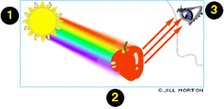
Vision begins when light rays are reflected off an object and enter the eyes through the cornea, the transparent outer covering of the eye. The cornea bends or refracts the rays that pass through a round hole called the pupil. The iris, or colored portion of the eye that surrounds the pupil, opens and closes (making the pupil bigger or smaller) to regulate the amount of light passing through. The light rays then pass through the lens, which actually changes shape so it can further bend the rays and focus them on the retina at the back of the eye. The retina is a thin layer of tissue at the back of the eye that contains millions of tiny light-sensing nerve cells called rods and cones, which are named for their distinct shapes. Cones are concentrated in the center of the retina, in an area called the macula. In bright light conditions, cones provide clear, sharp central vision and detect colors and fine details. Rods are located outside the macula and extend all the way to the outer edge of the retina. They provide peripheral or side vision. Rods also allow the eyes to detect motion and help us see in dim light and at night. These cells in the retina convert the light into electrical impulses. The optic nerve sends these impulses to the brain where an image is produced.The cornea is a transparent structure found in the very front of the eye that helps to focus incoming light. Situated behind the pupil is a colorless, transparent structure called the crystalline lens. A clear fluid called the aqueous humor fills the space between the cornea and the iris.Rods work at very low levels of light. We use these for night vision because only a few bits of light (photons) can activate a rod. Rods don't help with color vision, which is why at night, we see everything in a gray scale. The human eye has over 100 million rod cells.
Cones require a lot more light and they are used to see color. We have three types of cones: blue, green, and red. The human eye only has about 6 million cones. Many of these are packed into the fovea, a small pit in the back of the eye that helps with the sharpness or detail of images.
Other animals have different numbers of each cell type. Animals that have to see in the dark have many more rods than humans have.Another benefit to this layout is that the RPE can absorb scattered light. This means that your vision is a lot clearer. Light can also have damaging effects, so this set up also helps protect your rods and cones from unnecessary damage.
While there are many other reasons having the discs close to the RPE is helpful, we will only mention one more. Think about someone who is running a marathon. In order to keep muscles in the body working, the runner needs to eat special nutrients or molecules during the race. Rods and cones are similar, but instead of running, they are constantly sending signals. This requires the movement of lots of molecules, which they need to replenish to keep working. Because the RPE is right next to the discs, they can easily help reload photoreceptor cells and discs with the molecules they need to keep sending signals.
Now that we know how these photoreceptor cells work, how do we use them to see different colors?
We have three types of cones. If you look at the graph below, you can see each cone is able to detect a range of colors. Even though each cone is most sensitive to a specific color of light (where the line peaks), they also can detect other colors (shown by the stretch of each curve).
Since the three types of cones are commonly labeled by the color at which they are most sensitive (blue, green and red) you might think other colors are not possible. But it is the overlap of the cones and how the brain integrates the signals sent from them that allows us to see millions of colors. For example, the color yellow results from green and red cones being stimulated while the blue cones have no stimulation.

No comments:
Post a Comment